INDEX     BCH441 COURSE     HTML TUTORIAL     BIOINFORMATICS
A RasMol tutorial

Outline

Welcome to a short tutorial on RasMol.

Molecular Modeling
RasMol Installation
Downloading Coordinate Files
Stereo Vision
Representation
Selection
Color
H-Bonds
Measurements
Surfaces and Slabs
Saving and Printing
Scripts
Web Resources
Feedback

Molecular Modeling

Structure is a three-dimensional concept and it does not follow readily from any linear or tabular description of the protein. To study structure, it is necessary to develop an intuitive feeling of the spatial arrangement of typical protein features and this ultimately requires to experience proteins in 3-D. Roger Sayle's program RasMol is one of the most widely distributed programs for molecular modelling and viewing of small molecules, proteins and nucleic acids. It is compact, fast, versatile, rich with essential features and free. It is a command line driven program and can be scripted and the source code is available. For taking a quick look at things, or for teaching purposes, it is unsurpassed.

That is not to say that there are not alternatives (see ... link for an annotated list of available visualization software). Unfortunately, the most useful alternative to RasMol for teaching purposes, CHIME link, a web-browser plugin, only works with specific versions of IE, since the support for plugin interfaces was unexpectedly discontinued by Microsoft in the summer of 2001. MDL has since worked on updates, but CHIME will not run with IE 6. For the research communities, this is a great loss and there are still no good alternatives available to use with Web pages.

Rather than expect users to run a specific constellation of browsers and plugins, we will use short scripts in our Web pages, which can be copied and pasted into the command-line window of RasMol.

Molecular Graphics Examples

Molecular Graphics

Here are some examples for molecular graphics: an imunoglobulin FV fragment rendered with MOLSCRIPT, a Pleckstrin Homology domain rendered with Raster 3-D, and GFP, ray traced with POV-ray, with a glowing fluorophore in the centre. While rendering quality of such images is usually superior to a simple molecular viewer, such as RasMol, static images are of course not interactive.

RasMol Installation

Instructions for downloading and installing a current version of RasMol can be found at the OpenRasMol.org link Website. LINUX, Mac and many other binaries (even Windows) are available. The latest stable release is 2.7.1.1. Note that Microsoft Windows users need to install RasTop since Windows versions of Rasmol do not have copy and paste functionality.

You may consider configuring RasMol as a helper application for your Web browser. See the information page on Configuring Netscape ... linkat the University of Massachsetts RasMol site.

When you start the program, it will open two windows: a command line window and a viewer window. Type help into the command line window for a brief overview. Not all commands that work in this release are documented in the help feature. Here are three other sources of help (besides the help-file that came with your download:

  link   Quick Reference A PDF for a quick reference to important commands.
  link   RasMol Manual The online manual for RasMol 2.7.1.1
  link   Search ... Since RasMol is so widely distributed, using a Web-search engine is quite an effective way to get examples for the correct use of specific commands or to find how to solve certain non-obvious problems.

Downloading coordinate files

3-D molecular coordinate files for proteins and (some) nucleic acids are held at the Protein Data Bank (PDB) link. The following links will download coordinates to your disk:

link   2IMM Immunoglobulin VL: Variable Domain of the Light Chain (114 residues, 2.0 Å resolution).
link   1A6M Sperm-Whale Oxymyoglobin (151 residues, 1.0 Å resolution).

Stereo Vision

Being able to visualize and experience strucutre in 3-D is an essential skill, if you are at all serious about understanding the molecules of molecular biology.

Even though hardware devices exist that help in the three-dimensional perception of computer graphics images, for the serious structural biologist there is really no alternative to being able to fuse stereo pair images by looking at them. RasMol is an excellent tool to practice stereo vision and develop the skill. Stereo images consist of a left-eye and a right-eye view of the same object, with a slight rotation around the vertical axis (about 5 degrees). Your brain can accurately calculate depth from these two images, if they are presented to the right and left eye separately. This means you need to look at the two images and then fuse them into a single image - this happens when the left eye looks directly at the left image and the right eye at the right image.

In fact, it can also be the other way around and some people find convergent (cross-eyed) stereo viewing easier. I recommend the divergent (wall-eyed) viewing - not only because it is much more comfortable in my experience, but also because it is the default way in which stereo images in books and manuscripts are presented.

In order to visually fuse stereo image pairs, you need to override an ocular reflex that couples divergence and focussing, this is something that needs to be practiced for a while. Usually 5 to 10 minutes of practice twice daily for a week should be quite sufficient. It is not as hard as learning to ride a bicycle, but you need to practice regularily for some time, maybe 10 or 20 sessions of 3 to 5 minute over a period of a week or two. Once you have acquired the skill, it is really very comfortable and can be done effortlessly and for extended periods. You will enter a new world of molecular wonders !

Here are step by step instructions of how to practice stereo-viewing with RasMol.

Load a small protein into RasMol and display this as a simple backbone model.
Type set stereo -5 in the command line (the RasMol default is cross-eyed, thus the need to specify a negative rotation angle).
Resize the window, until two equivalent points on the protein are the same distance on the screen, as your eyes are apart (this is usually about 6 cm).
Touch your nose to the screen and look at the two images. They will be blurred and out of focus, but should appear as a three-dimensional object. Slowly rotating the protein helps.
Once you see the object in 3-D, try to move your head backwards slowly, until the structure comes into focus by itself. Do not voluntarily try to focus, since this will induce your eyes to converge and you will lose the 3-D effect. When you lose the 3-D effect, start over.
Practice this patiently, two times daily for some 3 to 5 minutes. Stop, when your head feels funny. Don't force yourself. It should take you about a week to master this, with regular training it will become very easy. And, the best thing is, you do not easily forget this skill. It is like riding a bicycle, equalizing pressure in your eustachian tubes while scuba diving, or circular breathing to play the didgeridoo: once you teach your body what to do, it remembers. And expands your horizon.

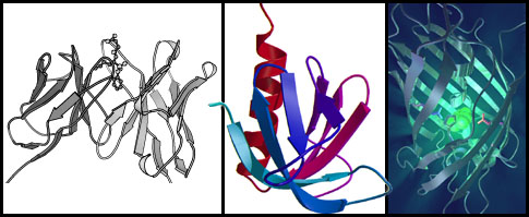
Below, there is a script to set up a stereo-view of 2IMM. Simply copy the script from the textarea and paste it into the RasMol command line window. The commands get executed line by line and it is easy to change parameters and arguments and see what effect this has.

A set of script commands for a simple view of 2IMM, suitable to practice stereo viewing.

2IMM in stereo

2IMM in stereo

This is approximately the result of the above commands in RasMol. The domain is shown as a backbone tube, connecting C-alpha atoms, the CDR regions are highlighted in light-green.

Here are some suggestions for stereo viewing mini-projects, so your practice sessions do not become monotonous. They are ordered from the most simple scenes to progressively more complicated molecules.

3D mini-project topics PDB source files
Study individual amino acids and memorize the spatial arrangement of the groups around the chiral centre (the C-alpha atom) in this L-amino acid. Alanine
Cysteine
Aspartate
Glutamate
Phenylalanine
Glycine
Histidine
Isoleucine
Lysine
Leucine
Methionine
Asparagine
Proline
Glutamine
Arginine
Serine
Threonine
Valine
Tryptophan
Tyrosine
Study (and remember) the correct chirality of the Threonine side-chain. Threonine
Download and study small molecule cofactors from HIC-UP. Some samples are linked here. ATP
Arachidonic acid
Beta-carotene
Biotin
Caffeine
FAD
FMN
Phycoerythrobilin
Testosterone
Study an alpha-helix. Concentrate on how the carbonyls are all oriented in the same direction. Since the carbonyl carries a significant negative dipole there is a large electrostatic dipole moment induced along the alpha helix. Memorize how this arrangement relates to the N- and C-terminus of the helix. Is it the N- or the C-terminus of the helix that lies in the strongly positive potential region of the helix dipole ? Where would a negatively charged residue (such as a phosphate group) find a binding site: at the beginning or the end of a helix ? Helix
Study a beta-sheet. Concentrate on how the alternating hydrogen bonds are formed between pairs of residuesin opposite direction. The example provided also has a cis-proline. Find it and study why the C-alpha atoms of the preceeding residue and of the proline are in cis, relative to the peptide bond ? Sheet
Display only the backbone, and the side chain atoms of Glu, Asp (color red) and Lys, Arg (color blue) residues in a protein. Pick out the salt-bridges in your structure.  
Download a DNA molecule. Study the phosphate backbone connections. Study and remember the arrangement of the 5' and 3' hydroxyl and the phosphate group. You should be able to discern the sequence of a DNA molecule from viewing the structure. You should also remember which way the helix turns: DNA is a right handed helix (except Z-DNA which is left-handed). B-DNA
A-DNA
Z-DNA
Study the catalytic triad in a serine protease. In this structure (one of the classics: bovine trypsin in complex with the pancreatic trypsin inhibitor, by Robert Huber and Johann Deisenhofer, 1982) the triad is serine 195, histidine 57 and aspartate 102. 2PTC.pdb
Study the ATP and t-RNA binding sites in a tRNA synthetase. Note the relative sizes of nucleotide structures and proteins. Pay special attention to the way the anticodon loop is tightly bound, as well as the acceptor stem. Using stereo, it should not be difficult to pick out the AMP molecule bound in this structure of E. coli glutaminyl tRNA synthetase right in the middle. 1EXD.pdb

Representation of Structures

RasMol allows a number of different representations of the molecule, they are accessible through the Menu, but they can also be entered through the command line and you need to know the equivalences, in order to construct scripts. Note that these commands also accept Boolean values as arguments i.e backbone off is a legal command while backbone on defaults to backbone 0. Note that the numerical parameters are in RasMol internal units of 1/250 Å.

Command line equivalences
Display Menu Command Line
Wireframe wireframe 0
Sticks wireframe 100
Backbone backbone 100
Spacefill cpk on or spacefill on
Ball & Stick wireframe 50
CPK 120
Cartoons wireframe 100 for strands
cartoon 380 for helices and strands

Note that the command cartoon is not documented in the help feature !

Note that water molecules do not show up in a wireframe view. In order to see single, non-bonded molecules, you have to select them (see below) and then display them as CPK spheres.

Script commands to show the two internal, structural water-molecules in 2IMM together with the residues in an eight Å radius around them. Hydrogen bonding atoms are colored.

Selection

RasMol has a very powerful command-line syntax to select parts of the protein structure. This is the most useful part of the program - since it makes real work far more efficient than having to use the mouse, once you understand the syntax and commands - but it is also one of the most confusing features of the priogram. It is a good idea to have a reference sheet handy. Load the structure for 1A6M.PDB for the next example. You can do this either with RasMols drag-and-drop feature or with the File - Open menu.

A set of script commands to demonstrate selections with a view of myoglobin in the 1A6M structure.

Here are the expression examples from the Quick Reference. Try some. Are you unsure sure how a specific atom is named? Just click it.

Examples for expressions
Command Meaning
* All atoms
cys Atoms in cysteines
hoh Atoms in water molecules
as? Atoms in asparagine or aspartic acid
*120 Atoms at residue 120 of all chains
*p Atoms in chain P
*.n? Nitrogen atoms
cys.sg Sulphur atoms in cysteine residues
ser70.c? Carbon atoms in serine-70
hem*p.fe Iron atoms in the Heme of chain P
*.*;A Atoms in alternate conformation A
*/4 All atoms in model 4 - this refers to multiple NMR-structure models in a single PDB file.

Note that you can combine commands with Boolean expressions - and, or and not ! For instance:
select (ser OR thr) AND NOT helix
is a valid selection. Note that AND in the logical sense is not used how you would colloquially use it: if you want both serine and threonine, you need to specify ser or thr. Writing ser and thr means that both ser as well as thr have to be true at the same time. This would select nothing.

Restricting is a concept very similar to selection, but it removes all parts that are not selected from the display. Try the following script. This also illustrates the useof the center command to center rotation to the center of gravity of a selection.

Script to focus on the heme group of myoglobin.

Color

Color can be entered either as a keyword, such as white or green, or as a triple of RGB values from 0 to 255 in square brackets e.g. [255,100,0] for orange. A number of predefined color schemes exist, that will color residues or atoms according to their properties. Important examples are

cpk     Color atoms by chemical type.
group    Color ramp from N-termuns (blue) to C-terminus (red). Note: if you cannot see the full color range in your protein, switch off display of heteroatoms and color again.
temperature    Color by B-value (disorder or mobility)

Other color choices are documented with the help function.

H-Bonds

H-bonds are drawn between the donating nitrogens and the accepting oxygens. Alternatively, in a backbone-only view, they can be drawn between C-alphas with set hbonds backbone. The commands for hydrogen bonds and disulfidebonds (ssbonds true) are used in a similar way.

H-bonds of the Myoglobin backbone.

Note that H-bonds are drawn for backbone atoms only - use monitor <atomnumber> <atomnumber> to draw lines between arbitrary atoms.

Measurements

Use the commands set picking distance, set picking angle and set picking torsion for measurements. The first, set picking distance, will get the distance between the next two atoms you pick with your mouse. The second will get the angle between the next three atoms and the third will calculate the dihedral angle between the next four atoms you pick. Type set picking off to return to the normal mode again. Try this: here are three amino acids from myoglobin. Get the PHI and the PSI angle for the residue in the middle !

Three aminoacids for picking and measuring backbone dihedral angles.
Verify you measurement with the following command select his24, show phipsi and confirm this with show ramprint. See the help section of the show command, help show, to find what other information is available.

Surfaces and Slabs

Rasmol can display van der Waals and solvent accessible surfaces and color them according to properties. Try the following for illustration:

The Van der Waals surface of the heme prosthetic group of myoglobin.

A "slab" or clipping plane allows you to see cross-sections of a molecule. Try this view:
A cross-section of Myoglobin shows the (red) heme-group packed into the hydrophobic interior (yellow).

You can move the slab forward and backward by pressing [control] [command] and the mouse on the Mac, [control] left-mouse-button on the PC. type help set slabmode and try out the different modes to view sections.

Saving and Printing

From the menu. Use write script <filename> to save a current view ! You can open, read and edit a script with any text-processor. Remember to save in text-only format when you are done.

Images can either be exported in a number of formats, or just copied (Edit - Copy from the Menubar) and then pasted e.g. into a MSWord document.

The File - Print command will print the current view.

Scripts

We have been using scripts all throughout this tutorial. But RasMol can also export its current state to a file, as a script. RasMol generated scripts are invaluable to determine the rotation and translation paramaters of a specific view. Use write script <filename> to have all the transformations and commands required to generate your current view dumped into a textfile. If no path is specified, the file will be in the directory from which RasMol was started. You can open this file with a text-processor and then copy and use or modify the required transformations. Other than that, the file writes the view on an atom-by-atom basis and is therefore not really suitable to study command syntax or as a source for editable scripts.

Web Resources

A collection of useful resources for RasMol on the Web.

WWW Resources
Site Description
PDB The repository of protein structure coordinates.
CATH Class, Architecture, Topology, Homology - a protein structure classification.
SCOP Structural Classification of Proteins.
HIC-Up Hetero Compound information database in Uppsala. Structures and data for the "other molecules", substrates, inhibitors, prosthetic groups etc. in the PDB.
Tutorial A RasMol tutorial by Gale Rhodes, University of Southern Maine.
RasMol The so called RasMol homepage at the University of Massachussets, by Eric Martz. Currently the site is pushing heavily for the author's CHIME implementation of a "protein explorer" which does not run natively under Unix operating systems. But there is still useful RasMol information too, especially a number of links to tutorials and scripts, if you search a little.

Feedback

Has this been helpful ? Inaccurate ? Confusing ? Do you have suggestions for improvement ? Do any of the topics need to be expanded ? Did you come accross an interesting resource on the Web ? I welcome all feedback to boris.steipe@utoronto.ca.


...
INDEX

...
BCH441 COURSE

...
HTML TUTORIAL

...
BIOINFORMATICS

U of T