BIN-SX-Small molecules
"Small Molecule" Structure"
(A small-molecule structure tutorial)
Abstract:
Creating with small molecule structures, finding complexes in the PDB that contain the molecule, and superimposing model and structure.
|
Objectives:
|
Outcomes:
|
Deliverables:
- Time management: Before you begin, estimate how long it will take you to complete this unit. Then, record in your course journal: the number of hours you estimated, the number of hours you worked on the unit, and the amount of time that passed between start and completion of this unit.
- Journal: Document your progress in your Course Journal. Some tasks may ask you to include specific items in your journal. Don't overlook these.
- Insights: If you find something particularly noteworthy about this unit, make a note in your insights! page.
Prerequisites:
You need the following preparation before beginning this unit. If you are not familiar with this material from courses you took previously, you need to prepare yourself from other information sources:
- Biomolecules: The molecules of life; nucleic acids and amino acids; the genetic code; protein folding; post-translational modifications and protein biochemistry; membrane proteins; biological function.
This unit builds on material covered in the following prerequisite units:
Contents
Evaluation
This learning unit can be evaluated for a maximum of 6 marks. If you want to submit tasks for this unit for credit you have the following options. If you have any questions about these options, discuss on the mailing list.
- Short report option
- 1. Create a new page on the student Wiki as a subpage of your User Page.
- 2. Visit the small-molecule selection page on the Student Wiki and choose one small molecule to work with.
- 3. Summarize what this molecule is. Draw the structure of the molecule in one of the molecular editors described in the unit and produce a SMILES string. Find a suitable PDB complex structure and superimpose your model in Chimera. Report on the quality of the superposition and create an informative stereo image that illustrates how your model is situated in the ligand binding site relative to the experimentally determined compound.
- 4. When you are done with everything, add the following category tag to the page:
[[Category:EVAL-BIN-SX-Small_molecules]]
- Do not change your submission page after this tag has been added. The page will be marked and the category tag will be removed by the instructor.
Contents
Task:
- Read the introductory notes on working with "small molecule" structure.
Modeling small molecules
"Small" molecules are solvent, ligands, substrates, products, prosthetic groups, drugs - in short, essentially everything that is not made by DNA-, RNA-polymerases or the ribosome. Whereas the biopolymers are still front and centre in our quest to understand molecular biology, small molecules are crucial for our quest to interact with the inventory of the cell, create useful products, or advance medicine.
A number of public repositories make small-molecule information available, such as PubChem at the NCBI, the ligand collection at the PDB, the ChEBI database at the European Bioinformatics Institute, the Canadian DrugBank, or the NCI database browser at the US National Cancer Institute. One general way to export topology information from these services is to use SMILES strings—a shorthand notation for the composition and topology of chemical compounds.
Task:
- Caffeine at PubChem
- Access PubChem.
- Enter "caffeine" as a search term in the Compound tab. A number of matches to this keyword search are returned.
- Click on the top hit - 1,3,7-Trimethylxanthine, the Caffeine molecule. Note that the page contains among other items:
- A 2D structural sketch;
- An idealized 3D structural conformer, for which you can download coordinates in several formats;
- The IUPAC name: 1,3,7-trimethylpurine-2,6-dione;
- The CAS identifier
58-08-2which is a unique identifier and can be used as a cross-reference ID; - The SMILES strings
CN1C=NC2=C1C(=O)N(C(=O)N2C)C; - ... and much more.
Task:
- Caffeine at DrugBank
- Access DrugBank.
- Enter "Caffeine" in the search form and. .
- Click on the hit to "Caffeine" itself. Note that the page contains among other items:
- A detailed description
- A 2D structural sketch with a link to 3D options;
- Synonyms, including the IUPAC name: 1,3,7-trimethylpurine-2,6-dione;
- ... and much more.
- Follow the link to the 3D options and note the options for downloading information, including the SMILES string and PDB formatted coordinates.
That's great, but let's sketch our own version of caffeine. Several versions of Peter Ertl's Java Molecular Editor (JME) are offered online, PubChem offers this functionality via its Sketcher tool and the PDB has a similar sketching tool on its ligand search page]
Task:
- Return to the PubChem homepage.
- Follow the link to Structure search (in the right hand menu).
- Click on the 3D conformer tab and on the Launch button to launch the molecular editor in its own window.
- Sketch the structure of caffeine. I find the editor quite intuitive but clicking on the Help button will give you a quick, structured overview. Make sure you define your double-bonds correctly.
- Export the SMILES string of your compound to your project folder.
Translating SMILES to structure
Chimera can translate SMILES strings to coordinates[1].
Task:
- Open Chimera.
- Select Tools → Structure Editing → Build Structure.
- In the Build Structure window, select the SMILES string button, paste the string from your file, and click Apply.
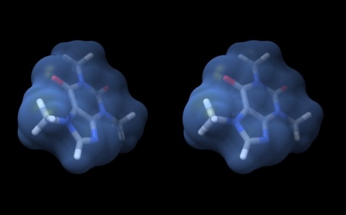
- The caffeine molecule will be generated and visualized in the graphics window. This is a "stick" representation.
- You can rotate it with your mouse, <command> drag to scale, <shift> drag to translate.
- Use the Actions → Atoms/Bonds → ball & stick or sphere menu items to change appearance.
- Use the Actions → Color → by element menu to change colors.
- Change the display back to stick and use Actions → Surface → show to add a solvent accessible surface. Choosing this command triggers the calculation of the surface, which is then available as an individually selectable object. However, with default parameters the surface appears a bit rough for this small molecule.
- Change the parameters of this solvent accessible surface:
- Select the surface with <control><click> (<control><left mouse button> on windows). A green contour line appears around selected items – it surrounds the surface in this case.
- Open the selection inspector by clicking on the tiny green icon in the lower-right corner of the window (It has a magnifying glass symbol which means "inspect" for Chimera, not "search").
- Select Inspect ...MSMS surface and change the Vertex density value to 50.0 - hit return.
- By default, the surface inherits the colour of the atoms it envelopes. To change the colour of the surface, use the Actions → Color → all options menu. Click the surfaces button to indicate that the color choice should be applied to the surface object (note what else you can apply color to...), then choose cornflower blue.
- Use the Actions → Surface → transparency → 50% menu to see atoms and bonds that are covered by the surface.
- To begin working with molecules in "true" 3D, choose Tools → Viewing Controls → Camera and select camera mode → wall-eye stereo. Also, use the Effects tab of the Viewing window, and check shadows off.
- Your structure should look about like what you see below.
Superposition
To investigate a small molecule structure variant in the context of a complex, we need to superimpose it with an existing ligand.
Task:
- Open the structure 3G6M in Chimera. This is one of the hits returned from the PDB serach for caffeine - a fungal chitinase for which caffeine is a potent inhibitor.
- Choose Select → Residue → CFF (CFF is th PDB three-letter code for this hetero compound), then Select → Invert (selected models), Actions → Atoms/Bonds → hide and Actions → Ribbon → hide to show only the caffeine molecules - there are two. Select the one with residue ID 1, and again Actions → Atoms/Bonds → hide. The remaining CFF molecule has residue ID 427.
To superimpose the structures, we can't use the standard "match" option, because that only works for protein or DNA molecules. Instead, we need to explicitly define matching pairs of atoms through Chimera's command line interface. The command line interface is a very powerful way to issue Chimera commands, but it has a bit of a learning curve since we need to use a precise model/residue/atom selection syntax.
- Visit the Chimera Help page on atom specification. Note how we specify models with a "#" sigil, residues with a ":" or "::" sigil, and atoms with an "@" sigil.
- Open the Chimera command line by clicking on the computer icon at the top left of the viewer window.
- The command we need is match, and we need to feed the command atoms in exactly the order of the pairs that the superposition algorithm should superimpose. To identify the atom numbers, we can hover over them with the mouse, or we can select the residue/atom and choose Actions → Label → name. If we superimpose the four nitrogen atoms, the correct command may be:
match #0@N3,N4,N1,N2 #1:427@N1,N3,N7,N9to superimpose the model we built from the SMILES string onto the structure - but the exact atom names in the model structure depend on how the SMILES string was written. - Note how the two structures virtually overlap - in this case, there are only very small coordinate differences because the conformational degrees of freedom are very much constrained in this xanthin hetrocycle. But there are differences nevertheless. One moleculae is an idealized structure, the other a structure that has been determined by a high-resolution experiment.
Self-evaluation
Notes
- ↑ There are also several online servers that translate SMILES strings to idealized structures, for example the online SMILES translation service at the NCI.
Further reading, links and resources
If in doubt, ask! If anything about this learning unit is not clear to you, do not proceed blindly but ask for clarification. Post your question on the course mailing list: others are likely to have similar problems. Or send an email to your instructor.
About ...
Author:
- Boris Steipe <boris.steipe@utoronto.ca>
Created:
- 2017-08-05
Modified:
- 2017-11-10
Version:
- 1.0
Version history:
- 1.0 First live version
- 0.1 First stub
![]() This copyrighted material is licensed under a Creative Commons Attribution 4.0 International License. Follow the link to learn more.
This copyrighted material is licensed under a Creative Commons Attribution 4.0 International License. Follow the link to learn more.
