Stereo vision practice
Stereo Vision Practice
This page contains scenes to practice stereo vision.
Here are example scenes to practice stereo viewing. However, they are static, and moving images help the eyes remain focused on the 3D effect. You could also refer to the BCH441 Stereo Vision Exam Questions.
Abstract Shapes
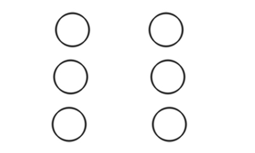
Three staggered circles. Side by side stereo image. These are three circles staggered from front (top) to back (bottom). There are no secondary visual depth-cues at all, such as perspective drawing, size changes or occlusions. Once you see the circles in stereo however, their spatial relationship should become vividly obvious.
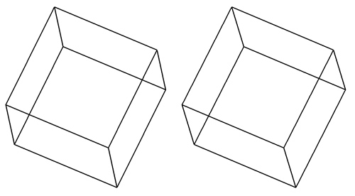
A cube. Side by side stereo image. This is a simple cube, drawn in orthographic projection and slightly rotated. Other than the pseudo-perspective angling of the lines going towards the back, there are no secondary visual depth-cues, such as occlusions or shading. This makes the 2D representation ambiguous regarding which face is closer to the viewer. If you look at only one of the images, you should be able to mentally invert the cube back-to front. In fact, when you concentrate on seeing the cube in a particular way, it becomes impossible to maintain this interpretation for more than about ten seconds or so: your visual system forces the switch and thus explores all of the ambiguous interpretations. Once you see the cube in stereo however, it becomes clear that the face further to the left is closer to you. Concentrating on the image in stereo enforces the correct interpretation, rather than causing a switch.
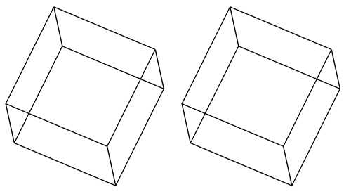
A flat cube. Not a stereo image! This is the left eye image from above in duplicate. If you look at this scene like a Side By Side stereo image, it appears flat, and the back-to-front switching phenomenon occurs.
Molecular Structures
An antibody Fab and its hapten
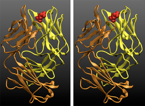
Side by side stereo image. The two chains of the Fab fragment of the phosphocholine-binding antibody McPC603 are shown in a cartoon representation, the light chain is in yellow (VL and CL domain), the two domains of the heavy chain are shown in orange (VH and CH1 - The CH2 and CH3 domains are part of the Fc fragment, not shown here). The bound hapten phosphorylcholine is shown in red in a surface representation.
Fold architecture in an antibody Fab
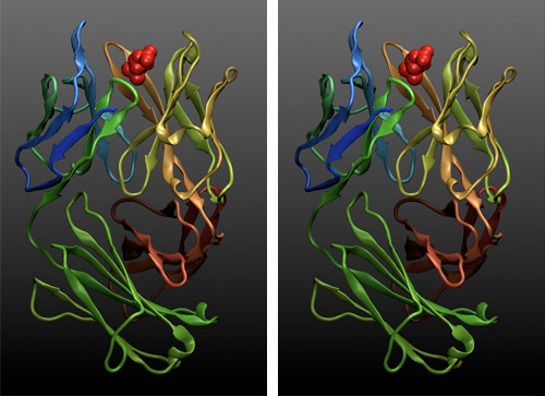
Side by side stereo image. The two chains of the structure have been colour-ramped according to their atom-index in the PDB file to highlight the overall fold of the four domains. These are so-called greek-key beta barrels: each domain comprises a two-sheet barrel with a hydrophobic core. The chain meanders first through half of the first sheet, crosses over the top of the domain, creates half of the other sheet, crosses back along the side, completes the first sheet, crosses under the domain and completes the second sheet. In stereo, it is easy to trace the chain architecture by eye.
The antigen binding site with bound phosphorylcholine
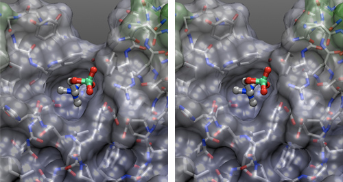
Side by side stereo image. A transparent solvent-accessible surface, lightly shaded by position has been laid over a licorice representation of the protein atoms. A ball-and-stick (CPK) representation was used for the hapten. Elements are color coded red for oxygen, silver for carbon, blue for nitrogen; phosphorous is green. In this representation we can discern element types and bonding topology of the structure, as well as the shape of the binding site that the protein conformation has generated.
A deep binding pocket accommodates the choline end of the hapten, the phosphate group is exposed to solvent. All molecular features of the hapten are discretely used in binding: the two oxygen atoms of a negatively charged aspartic acid can be seen at the bottom of the binding pocket, offsetting the positively charged trimethylammonium group of choline. Conversely, a positively charged arginine sidechain at the top, right, forms a salt-bridge with the negatively charged phosphate group. A tyrosine above the arginine hydrogen bonds one of the phosphate oxygens, while the hydrophobic sides of the binding pocket are formed by a tryptophan side chain (top) and a tyrosine sidechain (bottom).
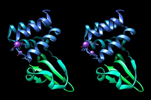
4IJE: Zaire ebolavirus VP35 interferon inhibitory domain mutant This simple structure is rendered as a ribbon model, with depth-cueing applied and a colour-ramp emphasizing the fold from N-C terminus.
Notes
Further reading and resources
| Westheimer (2011) Three-dimensional displays and stereo vision. Proc Biol Sci 278:2241-8. (pmid: 21490023) |
|
[ PubMed ] [ DOI ] Procedures for three-dimensional image reconstruction that are based on the optical and neural apparatus of human stereoscopic vision have to be designed to work in conjunction with it. The principal methods of implementing stereo displays are described. Properties of the human visual system are outlined as they relate to depth discrimination capabilities and achieving optimal performance in stereo tasks. The concept of depth rendition is introduced to define the change in the parameters of three-dimensional configurations for cases in which the physical disposition of the stereo camera with respect to the viewed object differs from that of the observer's eyes. |
