Difference between revisions of "Stereo Vision Exam Questions"
(→2004) |
(→2004) |
||
| Line 102: | Line 102: | ||
*'''Based on this figure, argue briefly why "G" is a conserved residue in the "<code>HIGH</code>" motif. ''' | *'''Based on this figure, argue briefly why "G" is a conserved residue in the "<code>HIGH</code>" motif. ''' | ||
</div> | </div> | ||
| + | <br> | ||
| + | <br> | ||
| + | |||
| + | ==2005== | ||
| + | |||
| + | Base specific recognition of canonically paired nucleotides can be mediated through specific recognition sites that contribute H-bond donors/acceptors or methyl groups. One example of such recognition sites for a particular basepair is sketched here: | ||
| + | |||
| + | [[Image:DNA_major-minor_groove.jpg|frame|none|In this sketch the major-groove recognition sites for a basepair are labelled A,B, C and D. ]] | ||
| + | |||
| + | <br> | ||
| + | |||
| + | The sketch corresponds to the situation in the stereo figure below. It was generated with the following RasMol commands to demonstrate the fold of the domain and several important sidechains on the recognition helix that interact with various parts of the DNA. | ||
| + | |||
| + | set background white | ||
| + | select all | ||
| + | wireframe off | ||
| + | restrict *c | ||
| + | color white | ||
| + | wireframe off | ||
| + | trace 90 | ||
| + | select (58,59,62,65-67) and *c | ||
| + | select (*.ca or sidechain) and selected | ||
| + | color [80,80,80] | ||
| + | wireframe 100 | ||
| + | select (5-11 and *a) or (3-9 and *b) | ||
| + | color [190,190,190] | ||
| + | wireframe 80 | ||
| + | select (*a or *b) and backbone | ||
| + | color [140,140,140] | ||
| + | set stereo -5 | ||
| + | |||
| + | <br> | ||
| + | |||
| + | [[Image:DNA_Elk-1_RecognitionHelix.jpg|frame|none|This divergent stereo-view shows the recognition helix of the Elk-1 transcription factor (pdb|1DUX) inserted into the major groove of a cognate oligonucleotide. ]] | ||
| + | |||
| + | <br> | ||
| + | |||
| + | <div style="padding: 5px; background: #DDDDDD; border:solid 1px #000000;"> | ||
| + | *'''Identify the "C" and "D" recognition interactions depicted in the sketch above. Note the one-letter code(s) for the amino acid type(s) that is/are involved in these interactions. ''' | ||
| + | </div> | ||
| + | |||
<br> | <br> | ||
<br> | <br> | ||
Revision as of 00:25, 12 December 2006
- Molecules are three-dimensional entities and stereo-vision of images on paper and on the screen is one of the most powerful, intuitive ways to appreciate that. We have practiced stero-vision in this course; here are a number of situations that require spatial awareness.
2002
- Write down the label of the tryptophan that is a conserved element of the hydrophobic core of this domain.
2002
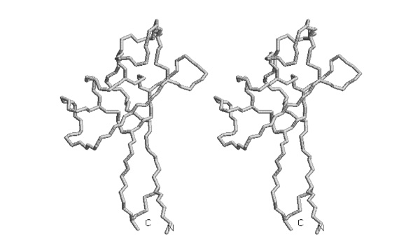
- Trace the backbone from the N-terminus to amino acid 52 with pencil or pen in one of the images.
I wouldn't ask this type of question any longer - the drawing task seems a bit convoluted for the actual skill it is supposed to test.
2003
As you know, a strong dipole moment is generated from the synergistic interactions of carbonyl groups in alpha helices. The carbonyls point towards the negative potential.
- Which three alpha helices are oriented best, so that their helix-dipole moment is aligned for favourable interactions with the ATP phosphate groups?
2003
The figure was generated with the following RasMol commands:
set background white
set stereo -5
select all
color white
restrict helix and backbone
wireframe 90
select glu,asp
color [80,80,80]
- Mark on this sheet the position of those Asp or Glu residues that are positioned to interact favourably with the helix dipole. If there are several plausible residues in a helix, mark the one closest to the correct terminus.
2003
- Trace the disulfide bonded cysteine sidechains in one of the stereoviews of this picture.
2004
- Number the cysteines from 1 to 8, from N- to C- terminus and determine the disulfide bonding topology of this protein. Write the disulfide bonded residue pairs into your exam booklet.
2004

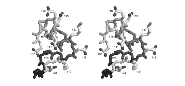
- List all pairs of residues that form salt-bridges according to this figure. Write the one-letter code for the negatively charged amino acid and its sequence number, then the same for the positively charged amino acid.
Comment
2004

HIGH" region is included here, its sequence is HVGT in this case. Carbons are shown in light grey, nitrogen atoms are darker, oxygen atoms are darkest grey and sulphur and phosphorous are black.
- Based on this figure, argue briefly why "G" is a conserved residue in the "
HIGH" motif.
2005
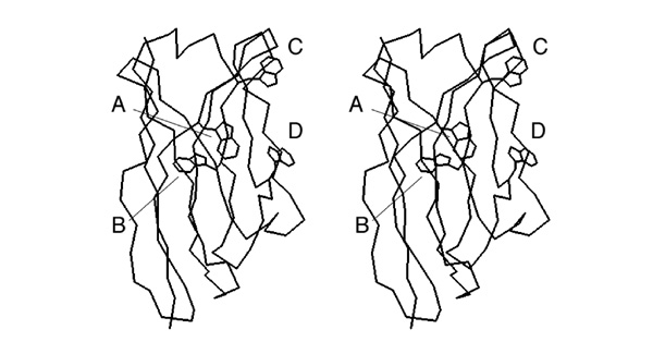
Base specific recognition of canonically paired nucleotides can be mediated through specific recognition sites that contribute H-bond donors/acceptors or methyl groups. One example of such recognition sites for a particular basepair is sketched here:
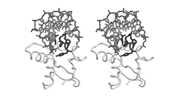
The sketch corresponds to the situation in the stereo figure below. It was generated with the following RasMol commands to demonstrate the fold of the domain and several important sidechains on the recognition helix that interact with various parts of the DNA.
set background white
select all
wireframe off
restrict *c
color white
wireframe off
trace 90
select (58,59,62,65-67) and *c
select (*.ca or sidechain) and selected
color [80,80,80]
wireframe 100
select (5-11 and *a) or (3-9 and *b)
color [190,190,190]
wireframe 80
select (*a or *b) and backbone
color [140,140,140]
set stereo -5
- Identify the "C" and "D" recognition interactions depicted in the sketch above. Note the one-letter code(s) for the amino acid type(s) that is/are involved in these interactions.


