Difference between revisions of "Stereo Vision Exam Questions"
Jump to navigation
Jump to search


(→2003) |
|||
| Line 5: | Line 5: | ||
;Molecules are three-dimensional entities and stereo-vision of images on paper and on the screen is one of the most powerful, intuitive ways to appreciate that. We have practiced stero-vision in this course; here are a number of situations that require spatial awareness. | ;Molecules are three-dimensional entities and stereo-vision of images on paper and on the screen is one of the most powerful, intuitive ways to appreciate that. We have practiced stero-vision in this course; here are a number of situations that require spatial awareness. | ||
</div> | </div> | ||
| − | | + | <br> |
| − | | + | <br> |
| + | |||
==2002== | ==2002== | ||
[[Image:Stereo_VH-domain.jpg|frame|none|This divergent stereo-view shows a trace of connected C<sup>α</sup> atoms of a protein domain (the VH domain of the anti-Fluorescein antibody 4-4-20, 4FAB.PDB) and a wireframe representation of all its tryptophan sidechains. ]] | [[Image:Stereo_VH-domain.jpg|frame|none|This divergent stereo-view shows a trace of connected C<sup>α</sup> atoms of a protein domain (the VH domain of the anti-Fluorescein antibody 4-4-20, 4FAB.PDB) and a wireframe representation of all its tryptophan sidechains. ]] | ||
| Line 14: | Line 15: | ||
;Write down the label of the tryptophan that is a conserved element of the hydrophobic core of this domain. | ;Write down the label of the tryptophan that is a conserved element of the hydrophobic core of this domain. | ||
</div> | </div> | ||
| − | + | <br> | |
| + | <br> | ||
==2002== | ==2002== | ||
| Line 25: | Line 27: | ||
<small>''I wouldn't ask this type of question any longer - the drawing task seems a bit convoluted for the actual skill it is supposed to test.</small> | <small>''I wouldn't ask this type of question any longer - the drawing task seems a bit convoluted for the actual skill it is supposed to test.</small> | ||
| + | <br> | ||
| + | <br> | ||
| + | |||
| + | ==2003== | ||
| + | [[Image:Stereo_ATP-binding_site.jpg|frame|none|This divergent stereo-view shows selected helices from 1GTR.pdb (backbone only, light grey) and the substrate ATP (dark grey). ]] | ||
| + | |||
| + | As you know, a strong dipole moment is generated from the synergistic interactions of carbonyl groups in alpha helices. The carbonyls point towards the negative potential. | ||
| + | |||
| + | <div style="padding: 5px; background: #DDDDDD; border:solid 1px #000000;"> | ||
| + | ;Which three alpha helices are oriented best, so that their helix-dipole moment is aligned for favourable interactions with the ATP phosphate groups? | ||
| + | </div> | ||
| + | <br> | ||
| + | <br> | ||
| + | |||
==2003== | ==2003== | ||
| Line 62: | Line 78: | ||
;Number the cysteines from 1 to 8, from N- to C- terminus and determine the disulfide bonding topology of this protein. Write the disulfide bonded residue pairs into your exam booklet. | ;Number the cysteines from 1 to 8, from N- to C- terminus and determine the disulfide bonding topology of this protein. Write the disulfide bonded residue pairs into your exam booklet. | ||
</div> | </div> | ||
| + | <br> | ||
| + | <br> | ||
Revision as of 15:08, 11 December 2006
- Molecules are three-dimensional entities and stereo-vision of images on paper and on the screen is one of the most powerful, intuitive ways to appreciate that. We have practiced stero-vision in this course; here are a number of situations that require spatial awareness.
2002

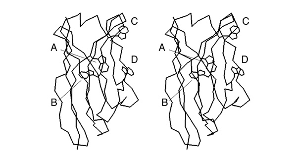
This divergent stereo-view shows a trace of connected Cα atoms of a protein domain (the VH domain of the anti-Fluorescein antibody 4-4-20, 4FAB.PDB) and a wireframe representation of all its tryptophan sidechains.
- Write down the label of the tryptophan that is a conserved element of the hydrophobic core of this domain.
2002

This divergent stereo-view shows a trace of connected Cα atoms of a Pleckstrin PH domain, 1PLS.PDB
- Trace the backbone from the N-terminus to amino acid 52 with pencil or pen in one of the images.
I wouldn't ask this type of question any longer - the drawing task seems a bit convoluted for the actual skill it is supposed to test.
2003

This divergent stereo-view shows selected helices from 1GTR.pdb (backbone only, light grey) and the substrate ATP (dark grey).
As you know, a strong dipole moment is generated from the synergistic interactions of carbonyl groups in alpha helices. The carbonyls point towards the negative potential.
- Which three alpha helices are oriented best, so that their helix-dipole moment is aligned for favourable interactions with the ATP phosphate groups?
2003

This divergent stereo-view shows selected helices from 1A2J.pdb (DsbA).
The figure was generated with the following RasMol commands:
set background white
set stereo -5
select all
color white
restrict helix and backbone
wireframe 90
select glu,asp
color [80,80,80]
- Mark on this sheet the position of those Asp or Glu residues that are positioned to interact favourably with the helix dipole. If there are several plausible residues in a helix, mark the one closest to the correct terminus.
2003

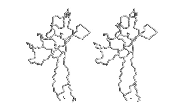
This divergent stereo-view shows a trace of connected backbone atoms – N, Cα, C and O – as well as the cysteine sidechains of the four disulfide bridges, of the pea defensin 1JKZ.pdb.
- Trace the disulfide bonded cysteine sidechains in one of the stereoviews of this picture.
2004

This divergent stereo-view shows a trace of connected backbone atoms – N, Cα and C – as well as the cysteine sidechains of the four disulfide bridges, of the pea defensin 1JKZ.pdb.
- Number the cysteines from 1 to 8, from N- to C- terminus and determine the disulfide bonding topology of this protein. Write the disulfide bonded residue pairs into your exam booklet.